Every human cell contains a nucleus, which houses genetic material in the form of genes. These genes hold the codes for all inherited traits and are organized on rod-like structures known as chromosomes.
Normally, a cell's nucleus has 23 chromosome pairs, with one set inherited from each parent. Down syndrome arises when there is a full or partial extra copy of chromosome 21.
The extra genetic material impacts developmental progress and leads to the traits seen in Down syndrome. Common physical features include low muscle tone, short stature, eyes that slant upwards, and a single deep crease across the palm center. However, individuals with Down syndrome are unique and may exhibit these traits to varying extents, or not at all.
According to the Centers for Disease Control and Prevention, approximately one in every 775 babies in the United States is born with Down syndrome, making Down syndrome the most common chromosomal condition. About 5,000 babies with Down syndrome are born in the United States each year.
-De Graaf, G., Buckley, F., & Skotko, B. (2024, May 3). People living with Down syndrome in the USA: Births and Population.
https://go.downsyndromepopulation.org/usa-factsheet
Throughout history, Down syndrome has been depicted in various forms of art, literature, and scientific texts. It wasn't until the late 19th century that John Langdon Down, an English physician, provided a comprehensive description of the condition. His landmark publication in 1866 earned him the title of "father" of the syndrome. Although the syndrome's characteristics were previously observed, Down was the first to categorize it as a distinct condition.
In recent years, advancements in medicine and science have deepened the understanding of Down syndrome. In 1959, French physician Jérôme Lejeune discovered that it is a chromosomal anomaly. He noted that individuals with Down syndrome possess 47 chromosomes in their cells, rather than the usual 46. This extra full or partial copy of chromosome 21 was identified as the cause of the syndrome's characteristics. In 2000, an international team of researchers cataloged approximately 329 genes on chromosome 21, which has led to significant progress in Down syndrome research.
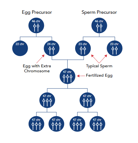
Down syndrome typically arises from a cell division error known as "nondisjunction." This error leads to an embryo having three copies of chromosome 21, rather than the standard two. This occurs when a pair of 21st chromosomes in either the sperm or the egg does not separate properly before or at the time of conception. As a result, the additional chromosome is duplicated in every cell of the body. The most common form of Down syndrome, accounting for 95% of cases, is referred to as trisomy 21.
Mosaicism, or mosaic Down syndrome, is identified when a person has a mix of two types of cells: some with the typical 46 chromosomes and others with 47. The cells with 47 chromosomes have an additional chromosome 21.
Mosaicism is the rarest form of Down syndrome, representing about 2% of all Down syndrome cases (Facts about Down syndrome, 2021). Studies suggest that individuals with mosaic Down syndrome might exhibit fewer characteristics of Down syndrome compared to those with other forms. However, it's not possible to make broad generalizations due to the diverse range of abilities seen in people with Down syndrome.
In translocation, accounting for approximately 3% of Down syndrome cases, the total chromosome count in cells remains 46. However, an extra full or partial copy of chromosome 21 becomes attached to another chromosome, often chromosome 14. This additional genetic material is responsible for the characteristics associated with Down syndrome.
Regardless of the specific type of Down syndrome a person may have, all individuals with Down syndrome possess an extra, critical segment of chromosome 21 in some or all of their cells.
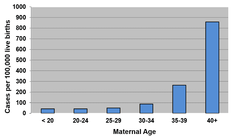
The origins of the extra full or partial chromosome remain unknown. Age is the only factor consistently linked to an increased likelihood of having a child with Down syndrome due to nondisjunction or mosaicism. Nevertheless, because younger women have higher birth rates, 51% of children with Down syndrome are born to women under the age of 35 (De Graaf et al., 2022).
No definitive scientific evidence suggests that environmental factors or parental activities before or during pregnancy cause Down syndrome.
The extra partial or full copy of chromosome 21 that leads to Down syndrome can come from either parent. About 5% of cases are attributed to the father.
Down syndrome affects individuals across all races and economic backgrounds, with older women having a higher likelihood of giving birth to a child with Down syndrome. The probability for a 35-year-old woman is approximately 1 in 350, which increases to 1 in 100 by the age of 40, and by 45, the chance is about 1 in 30. The risk of translocation does not appear to be associated with the mother's, or birthing parent's, age.
As more couples choose to have children later in life, the occurrence of Down syndrome conceptions may rise. Consequently, genetic counseling for prospective parents is gaining importance. However, many physicians lack complete information to guide their patients regarding Down syndrome incidence, diagnostic advancements, and care and treatment protocols for infants diagnosed with Down syndrome.
All three types of Down syndrome are genetic disorders, yet only 1% of cases have a hereditary element where the condition is passed from parent to child. Heredity does not play a role in trisomy 21 (nondisjunction) or mosaicism. Nonetheless, approximately one-third of Down syndrome cases caused by translocation do have a hereditary component, which represents about 1% of all instances of Down syndrome (Facts about Down syndrome, 2021).
Parental age does not appear to influence the risk of translocation. While most occurrences are random, in roughly one-third of the cases, a parent carries a translocated chromosome.
After a parent has had a child with trisomy 21, either through nondisjunction or translocation, the likelihood of having another child with trisomy 21 is estimated to be 1 in 100 until the age of 40.
The recurrence risk for translocation depends on the parent who carries the genetic alteration: approximately 3% if the father is the carrier, and between 10-15% if the mother is the carrier. Genetic counseling can help identify the source of the translocation.
Two categories of tests for Down syndrome are available before birth: screening and diagnostic tests. Prenatal screenings estimate the likelihood of the fetus having Down syndrome, providing a probability rather than a certainty. Diagnostic tests, however, can almost definitively diagnose Down syndrome with nearly 100% accuracy.
A wide range of prenatal screening tests are available to expectant parents. These typically include a blood test and an ultrasound. The blood tests, also known as serum screening tests, assess the levels of certain substances in the parent's blood. These levels, along with the parent's age, help estimate the risk of Down syndrome. Often, these tests are combined with a detailed sonogram to look for "markers" associated with Down syndrome. Newer advanced screens can detect fetal chromosomal material in the maternal blood, offering high accuracy without being invasive like the diagnostic tests that follow. However, these screens cannot provide a definitive diagnosis of Down syndrome. Both prenatal screening and diagnostic tests are now commonly offered to parents of all ages.
For a definitive prenatal diagnosis of Down syndrome, the available procedures are chorionic villus sampling (CVS) and amniocentesis. These carry a small risk of miscarriage, up to 1%, but are nearly 100% accurate. Amniocentesis is typically done in the second trimester, between 15 and 20 weeks of gestation, while CVS is performed in the first trimester, between 11 and 14 weeks (Chorionic Villus Sampling, 2020).
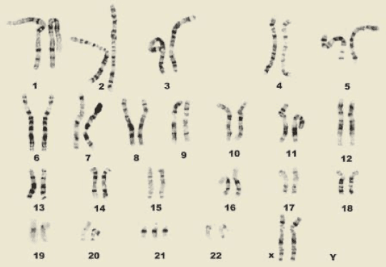
Down syndrome is typically recognized at birth by certain physical characteristics: low muscle tone, a single deep crease across the palm, a somewhat flattened facial profile, and eyes that slant upwards. However, since these traits can also occur in babies without Down syndrome, a chromosomal analysis known as a karyotype is performed to confirm the diagnosis. This involves drawing a blood sample to examine the baby's cells, photographing the chromosomes, and grouping them by size, number, and shape. Through the karyotype, doctors can identify Down syndrome. Additionally, a genetic test called fluorescence in situ hybridization (FISH) can quickly confirm a diagnosis by visualizing and mapping an individual's genetic material in their cells.


People with Down syndrome are increasingly becoming part of various community organizations, including schools, healthcare systems, workplaces, and social as well as recreational activities. They exhibit a range of cognitive delays, which can be very mild to severe, although most experience mild to moderate delays.
Thanks to medical advancements, the life expectancy of individuals with Down syndrome has significantly increased. In 1910, the life expectancy for children with Down syndrome was around nine years. The advent of antibiotics extended this to an average of 19 or 20 years. Currently, due to improvements in clinical treatments, especially heart surgery, up to 80% of adults with Down syndrome now live to 60 years or more. Consequently, there is a growing interaction between Americans and individuals with Down syndrome, highlighting the importance of public education and acceptance.